
Revealing the structure of axons
Biology
Axons, the threadlike part of a nerve cell that conducts impulses, are both flexible and strong, which makes them a mystery in the eyes of biologists. Recent studies have shown that under the axonal membrane, rings composed of actin filaments give the structure its flexibility. But those studies had not been able to define the precise architecture of these rings. By combining two microscopy techniques, optical and electronic, researchers at the Institut de Neurophysiopathologie (CNRS/Aix-Marseille Université) and the Institut de Myologie (INSERM/Sorbonne Université) have now managed to observe these rings at the molecular scale. They are formed of long braided actin filaments, braided like a Christmas wreath. This work, which brings key new insights into our understanding of axonal architecture, was published on 20 December 2019 in Nature Communications.
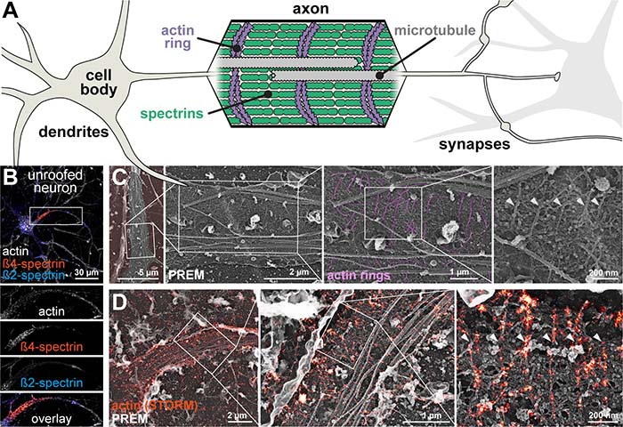
©Christophe Leterrier & Stéphane Vassilopoulos/CC
Bibliography
Ultrastructure of the axonal periodic scaffold reveals a braid-like organization of actin rings. Stéphane Vassilopoulos, Solène Gibaud, Angélique Jimenez, Ghislaine Caillol et Christophe Leterrier. Nature Communications, decembre 20, 2019. DOI : 10.1038/s41467-019-13835-6
Contact
Christophe Leterrier
CNRS Researcher