
A methodological leap in the exploration of memory
Biology
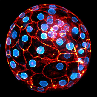
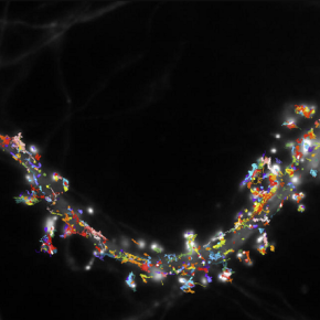
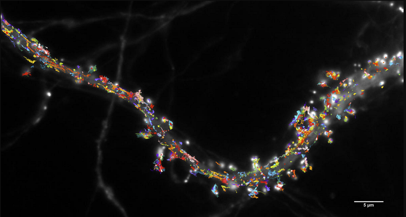
Neurons communicate with each other across synapses, areas of close contact where neurotransmitter molecules released from one neuron act on receptors embedded in the membrane of the opposite neuron. Previous research conducted by the team of Daniel Choquet, researcher at the CNRS and Director of the Interdisciplinary Institute for Neurosciences (CNRS/University of Bordeaux) had discovered that these receptors are not stationary, but instead move constantly in the membrane. The same scientists suggested and indirectly demonstrated that this movement modifies the number of receptors in a synapse at a given time to modulate the effectiveness of synaptic transmission and, as a result, certain types of learning and memory.1 Until now, however, it was not possible to observe receptor mobility in situ, in situations more natural than neuron cultures. This has now been achieved: thanks to the development of a comprehensive 'toolbox', scientists have been able to establish that this mobility exists in intact brain tissue, and that it is indispensable for certain types of memory, such as the contextual fear memory tested in this study. This 'toolbox' consists of a new animal model, improved high-resolution imaging technology and techniques for labelling and controlling receptor dynamics. It will allow the study of any region of the brain in addition to the hippocampus, can be transposed to other types of receptors, and will be used by the team to study the possible role of receptor mobility in intellectual disabilities and autism spectrum disorders.
This research was supported by an ERC Advanced grant awarded to Daniel Choquet.
To find out more, read this interview with Daniel Choquet on CNRS News: How imaging is revolutionising biology (October 2021).
© Benjamin Compans et Daniel Choquet / IINS / CNRS-Université de Bordeaux
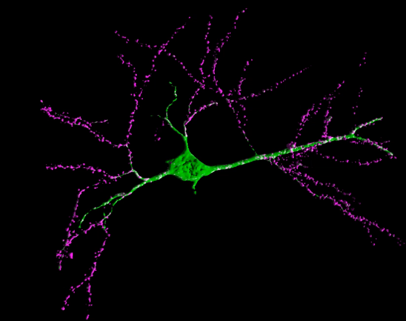
© Angela Getz, Mathieu Ducros, Daniel Choquet / IINS et BIC / CNRS-Université de Bordeaux-Inserm.
- 1Synaptic receptor mobility: discovery of a new mechanism for controlling memory (13 September 2017) https://www.cnrs.fr/en/synaptic-receptor-mobility-discovery-new-mechanism-controlling-memory
Bibliography
High-resolution imaging and manipulation of endogenous AMPA receptor surface mobility during synaptic plasticity and learning, Angela M. Getz, Mathieu Ducros, Christelle Breillat, Aurélie Lampin-Saint-Amaux, Sophie Daburon, Urielle François, Agata Nowacka, Mónica Fernández-Monreal, Eric Hosy, Frédéric Lanore, Hanna L. Zieger, Matthieu Sainlos, Yann Humeau & Daniel Choquet. Science Advances, 27 July 2022. DOI: 10.1126/sciadv.abm5298